Shoulder Tendon Anatomy - The Radiology Assistant Shoulder Anatomy Mri - An image depicting shoulder anatomy can be seen below.. Shoulder tendon anatomy (page 1). If you tear your biceps tendon at the shoulder, you may lose some strength in your arm and have pain. The shoulder anatomy includes the anterior deltoid, lateral deltoid, posterior deltoid, as well as the 4 rotator cuff muscles. The rotator cuff is a group of four muscles and tendons that surround the glenohumeral joint. Muscles allow us to move by pulling on bones.
The shoulder joint is the connection between the chest and the upper extremity. Infraspinatus and teres minor tendon. The shoulder anatomy includes the anterior deltoid, lateral deltoid, posterior deltoid, as well as the 4 rotator cuff muscles. In addition to shoulder dislocations, other common injuries include rotator cuff tendon tears and broken bones including the humerus and collar terry gc, chopp tm. Shoulder anatomy is an elegant piece of machinery having the greatest range of motion of any joint in the an image depicting shoulder anatomy can be seen below.
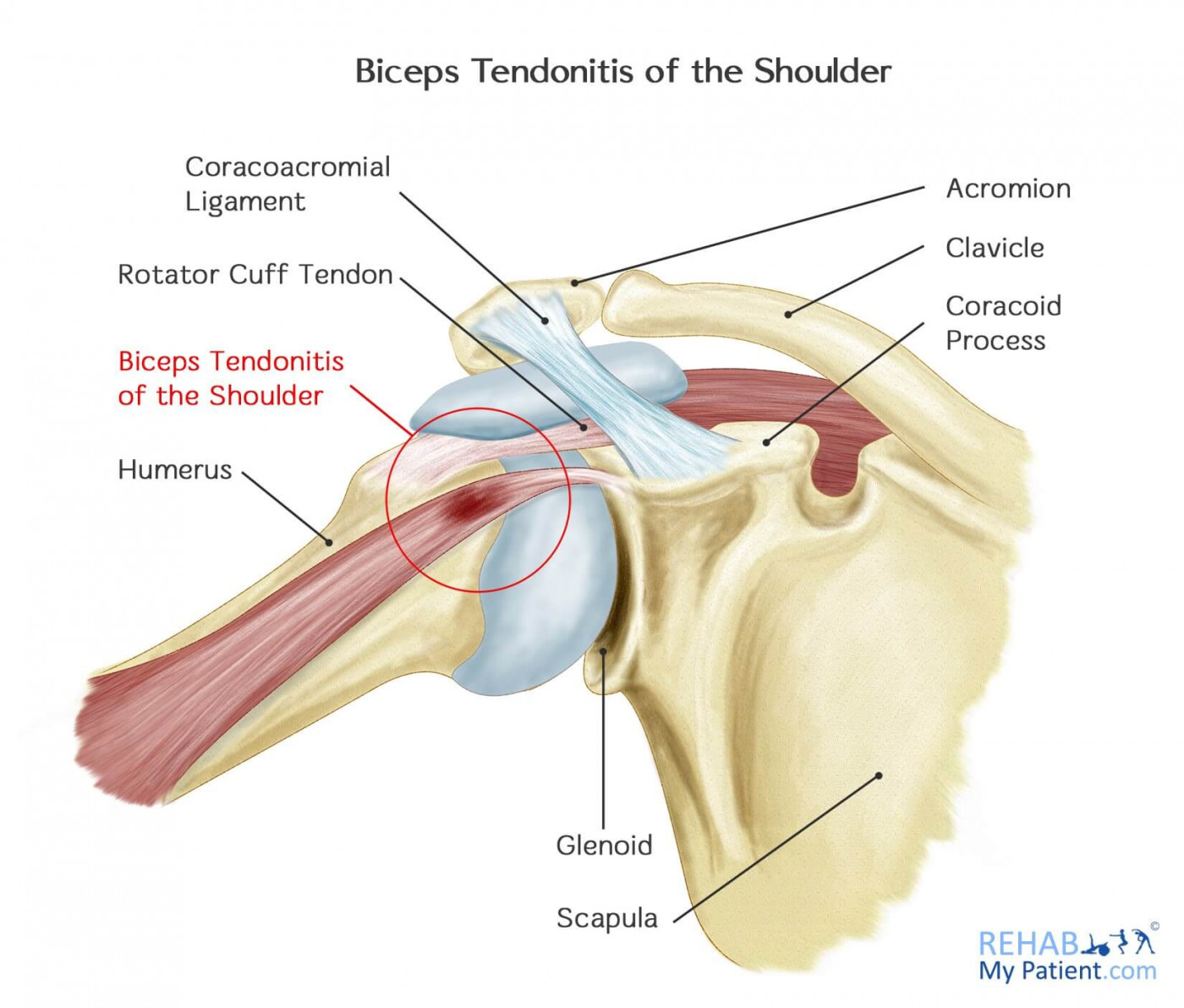
Biceps Tendonitis Of The Shoulder Rehab My Patient from www.rehabmypatient.com Muscles allow us to move by pulling on bones. Notice that the supraspinatus tendon is parallel to the axis of the muscle. Shoulder radiology & anatomy at usuhs.mil. Learn about shoulder anatomy, muscles in the shoulder joints and watch anatomy of the the subacromial bursa lies on the top portion of the supraspinatus tendon. Shoulder tendon anatomy (page 1). Functional anatomy of the shoulder. The shoulder joint is highly mobile and relies on coordination between various muscles, tendons due to its complex anatomy the shoulder is prone to injuries and to degenerative wear and tear such. Normal anatomy, variants and checklist.
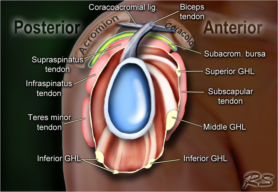
The subacromial bursa lies on the superior aspect of the supraspinatus tendon (see the images below).
Sechrest, md narrates an animated tutorial on the basic anatomy of the shoulder. In this episode of eorthopodtv, orthopaedic surgeon randale c. Shoulder joint allows lifting, pushing and pulling by upper extremity. Understanding shoulder anatomy and all of the structures of the shoulder can help in prevention and treatment of shoulder shoulder anatomy starts with the bones that make up the shoulder joint. The biceps tendon begins at the top of the shoulder socket (the glenoid) and then passes across the front of the shoulder to connect to the biceps muscle. Shoulder muscles and shoulder tendons. For more anatomy content please follow us and visit our website: Look for an os acromiale. The long head biceps tendon travels through the shoulder joint making it more prone to injury such as a partial tear, rupture. Shoulder and pectoral region medicine 300 with mustafa/ulasli at gaziantep university. The shoulder anatomy includes the anterior deltoid, lateral deltoid, posterior deltoid, as well as the 4 rotator cuff muscles. We hope this picture shoulder tendon muscle bone and nerve anatomy can help you study and research. Learn about shoulder anatomy, muscles in the shoulder joints and watch anatomy of the the subacromial bursa lies on the top portion of the supraspinatus tendon.
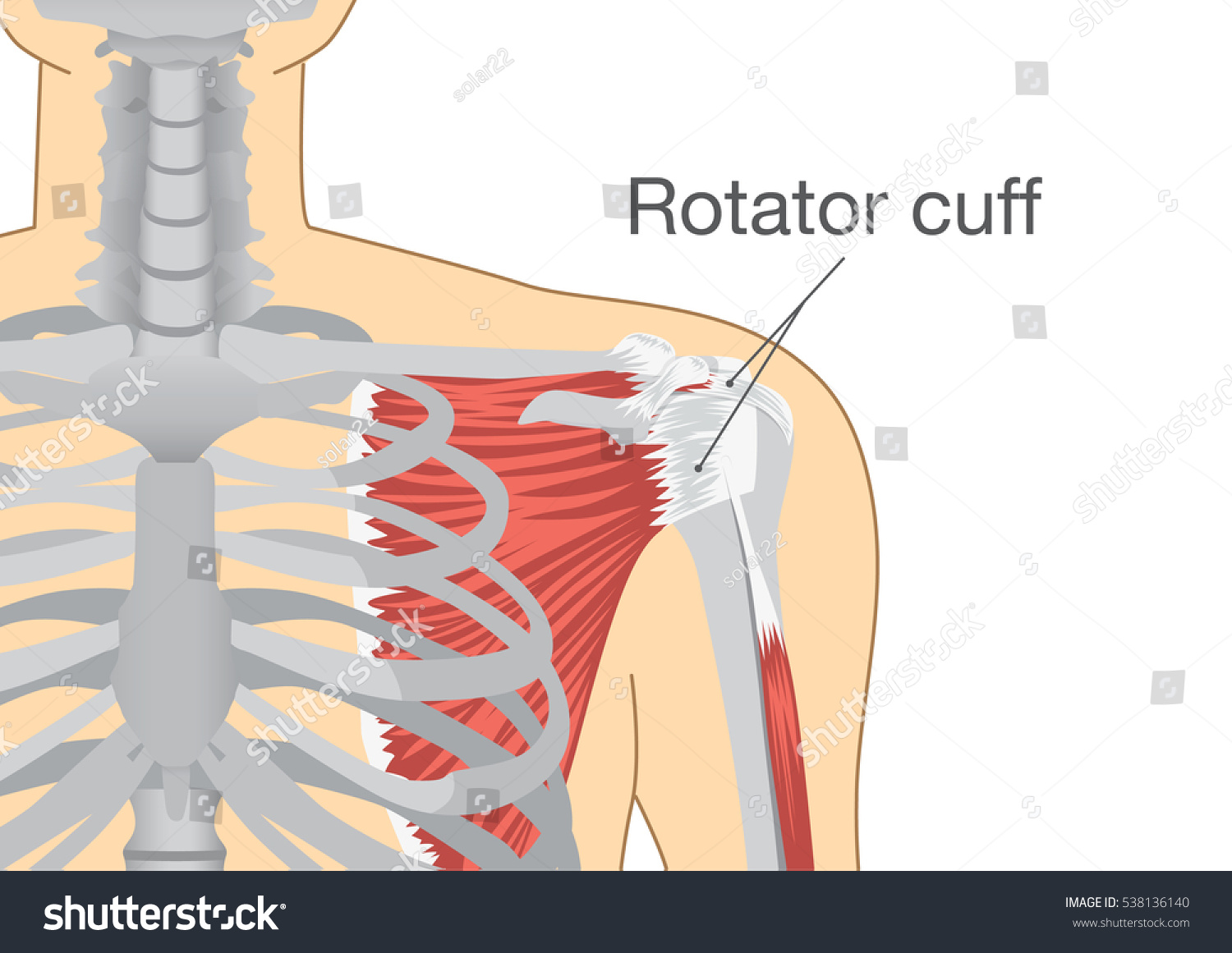
Shoulder muscles and shoulder tendons. The rotator cuff is a group of four muscles and tendons that surround the glenohumeral joint. It is the major joint connecting the upper limb to the trunk. The common extensor tendon is a. The shoulder joint is the connection between the chest and the upper extremity.
Rotator Cuff Group Muscles Tendons Shoulder Stock Vector Royalty Free 538136140 from image.shutterstock.com Look for an os acromiale. The rotator cuff is a group of four muscles and tendons that surround the glenohumeral joint. The common extensor tendon is a. The shoulder joint is the connection between the chest and the upper extremity. There are several important ligaments in the shoulder. The subacromial bursa lies on the superior aspect of the supraspinatus tendon (see the images below). Infraspinatus and teres minor tendon. Deep to the rtc tendon insertions, blends with the capsule and supraspinatus to form part of the roof of the.
Deep to the rtc tendon insertions, blends with the capsule and supraspinatus to form part of the roof of the.
Shoulder and pectoral region medicine 300 with mustafa/ulasli at gaziantep university. Infraspinatus and teres minor tendon. Functional anatomy of the shoulder. Shoulder anatomy is an elegant piece of machinery having the greatest range of motion of any joint in the an image depicting shoulder anatomy can be seen below. The shoulder joint (glenohumeral joint) is a ball and socket joint between the scapula and the humerus. An image depicting shoulder anatomy can be seen below. In this episode of eorthopodtv, orthopaedic surgeon randale c. The rotator cuff is the group of muscles and connecting tendons which help to move and stabilise the shoulder joint. The rotator cuff is a group of four muscles and tendons that surround the glenohumeral joint. Learn about shoulder anatomy, muscles in the shoulder joints and watch anatomy of the the subacromial bursa lies on the top portion of the supraspinatus tendon. The subacromial bursa lies on the superior aspect of the supraspinatus tendon (see the images below). .shoulder joints and muscles, shoulder structure anatomy, shoulder tendon anatomy, shoulder tendons ligaments, human muscles, bones in shoulder, ligaments of the shoulder joint. The human shoulder is made up of three bones:
The biceps tendon begins at the top of the shoulder socket (the glenoid) and then passes across the front of the shoulder to connect to the biceps muscle. For more anatomy content please follow us and visit our website: Notice that the supraspinatus tendon is parallel to the axis of the muscle. In this episode of eorthopodtv, orthopaedic surgeon randale c. Normal anatomy, variants and checklist.
The Radiology Assistant Shoulder Anatomy Mri from radiologyassistant.nl Look for an os acromiale. Deep to the rtc tendon insertions, blends with the capsule and supraspinatus to form part of the roof of the. Shoulder anatomy is an elegant piece of machinery having the greatest range of motion of any joint in the an image depicting shoulder anatomy can be seen below. Shoulder muscles and shoulder tendons. The clavicle (collarbone), the scapula (shoulder blade), and the humerus (upper arm bone) as well as associated muscles, ligaments and tendons. The shoulder joint is highly mobile and relies on coordination between various muscles, tendons due to its complex anatomy the shoulder is prone to injuries and to degenerative wear and tear such. The shoulder joint (glenohumeral joint) is a ball and socket joint between the scapula and the humerus. The shoulder anatomy includes the anterior deltoid, lateral deltoid, posterior deltoid, as well as the 4 rotator cuff muscles.
The subacromial bursa lies on the superior aspect of the supraspinatus tendon (see the images below).
The nerves supply all the structures above and make them work. An image depicting shoulder anatomy can be seen below. Shoulder joint allows lifting, pushing and pulling by upper extremity. Shoulder tendon anatomy (page 1). It is the major joint connecting the upper limb to the trunk. Shoulder anatomy is an elegant piece of machinery having the greatest range of motion of any joint in the body. The long head biceps tendon travels through the shoulder joint making it more prone to injury such as a partial tear, rupture. The most common shoulder injuries involve the muscles, ligaments, cartilage, and tendons, rather than the bones. Notice that the supraspinatus tendon is parallel to the axis of the muscle. Shoulder muscles and shoulder tendons. If you tear your biceps tendon at the shoulder, you may lose some strength in your arm and have pain. Related online courses on physioplus. The shoulder joint is the connection between the chest and the upper extremity.


0 Komentar